|
Welcome to a VIA Online Learning Tutorial: This site has been developed to provide learning materials to enable you to create X-ray exposure charts.
Select Chapter:
1. INTRODUCTION
2. STARTING
3. CREATING
4. OPTIMISED [print this page]

Optimised Exposure Charts
The previous sections described how to construct a general purpose kVp-variable exposure chart. It is a good idea to learn how to adapt charts to produce optimal exposures of the many different body regions encountered in small animal radiography.
Consider radiographing finely detailed structures, such as:
• extremities and dentition of cats and dogs
• feline thoraxes, and
• the entire bodies of birds and small exotic species.
As well, for X-ray machines that have an extended kVp range (at least up to 125) and high mA (at least 300), additional charts can be constructed to produce superior radiographs of canine thoracic and skeletal structures.
Detail radiography
Detail radiographs can be made using almost any X-ray machine regardless of its level of sophistication. This technique relies on the availability of receptor systems designed to produce images with increased detail.
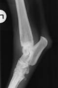 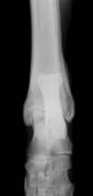
Fig 1: Canine tibio-tarsal joint radiographed using a detail receptor system.
Intensifying screens optimised for detail are slow systems, generally rated at a speed of 100-200 when used with double-emulsion X-ray film. They are even slower if used with single-emulsion film.
The following example chart was created for radiographing the extremities of dogs and cats using a detail receptor system. The mAs was constant, at 12 mAs.
This type of radiography doesn't include a grid, because of the small tissue thicknesses involved (generally 2-9cm) but you will need to use 10-20mAs to overcome the slow receptor speed.
To construct a kVp-variable chart optimised for detail radiography, select the structure for which the chart is to be built an extremity of cat or dog, cat thorax, bird or small exotic animal then find an animal on which the depth of the structure measures about 4-6cm and follow the instructions for a standard kVp-variable chart for spinal radiography, leaving out the grid and calculating exposure factors for tissue depths for 2-9cm only. Select 10-15mAs and 45-55kVp as the initial settings for the first test exposure.
High-kVp radiography
High-kVp radiography is ideally suited to producing diagnostic images of the lungs because high X-ray energies lengthen the radiographic grey-scale while decreasing the number of photons absorbed by osseous structures.
Reducing the grey-scale improves the clarity of pulmonary tissue by compensating for gas-induced increases in radiographic contrast.
As well, decreasing the amount of photons absorbed by osseous structures reduces the intrusion of images of the ribs into pulmonary radiographs.
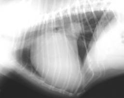 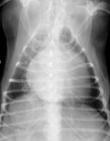
Fig 2: Radiographs taken using exposure factors optimised for the thorax.
By taking advantage of these two effects, high-kVp thoracic radiography improves the radiographers ability to record even small changes in the opacity of the lungs.
Because of the relationship between kVp and mAs an exposure chart designed for high kVp will require a low mAs.
The following example chart was optimised for high-kVp thoracic radiography by starting with 100kVp and 1.1 mAs:

Low-kVp radiography
Low-kVp radiography, which is the corollary to high-kVp radiography, is particularly suited to producing images of the vertebrae, skull and hips.
Selecting low beam energies causes increased photoelectric absorption of X-ray photons in tissue, which dramatically shortens the grey-scale of the resulting radiograph, producing high-contrast images.
Such an exposure strategy is useful for radiographing osseous structures, which benefit from enhanced tissue contrast.
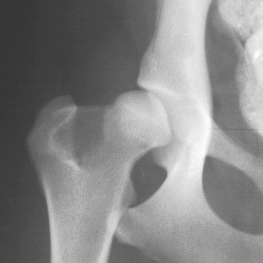
Fig 3. Close up, canine coxofemoral joint
Because of the relationship between kVp and mAs, an exposure chart designed for low kVp will require high mAs.
Anyone contemplating low-kVp-high-mAs radiography needs to be aware of the tube rating and overload limits of their X-ray machine. Superior tissue contrast is available for those whose equipment can take advantage of this technique.
The following example chart was created using the kVp-variable method for standard radiography, then optimising for low-kVp hip radiography. The mAs was a constant 43 mAs.
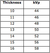
To construct a kVp-variable chart optimised for low-kVp radiography, follow the instructions for a standard kVp-variable chart for spinal radiography, substituting 40-50kVp and 30-60mAs as the initial settings for the first test exposure.
<< Previous chapter "Creating"
|