Welcome to a VIA Online Learning Tutorial: This site has been developed to provide learning materials to assist you to make complete and comprehensive radiographic examinations of animals.
This article has been prepared by Leanne Fitzsimmons. Leanne was the practice manager of Veterinary Imaging Associates, a practice of specialist veterinary radiologists who evaluate hips and elbows for for the AVA/ANKC certification scheme. They also provide hip and elbow dysplasia advice to interested individuals and are PennHIP® licensed. Each radiograph sent to Veterinary Imaging Associates for hip or elbow scoring is viewed by two radiologists before a score is finalised. For further information please call on 1300 300 892 or email on via@online-vets.com
Canine Hip Dysplasia (CHD) is the most common heritable orthopaedic problem seen in dogs. It affects virtually all breeds of dogs, but is especially problematic in large and giant breeds. CHD develops into a degenerative condition (osteoarthritis) of the hip joints. Conventionally, CHD is diagnosed radiographically by the presence of degenerative changes and/or subluxation of the hip joint[s]. The role of subluxation is crucial in the development of CHD but is often camouflaged on the most commonly used radiographic projection of the hips. Radiographic evidence of osteoarthritis confirms secondary changes of CHD, characterised by periarticular osteophyte formation (bone spurs), signs of increased subchondral bony opacity and bony remodelling.
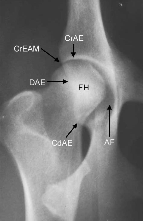
The current system used for scoring radiographs for hip dysplasia in Australia is based the system devised and used by the BVA/KC. There are nine criteria (see diagram) to be evaluated. Scores between 0 and 6 are allocated for all criteria, except the caudal acetabular edge, for which the maximum score is 5. Higher scores indicate greater degrees of radiographic abnormality. The scores for the right and left joints are added to give a total hip score but the status of the worst individual hip is used for grading purposes where grading systems [as in Europe] are used. The anatomical landmarks used for scoring are illustrated below.
[click to enlarge]
For a number of reasons it is not possible to rigidly standardise the information provided by the radiographic image of a complex structure such as the hip joint. Some of these reasons are:
• conformation; there is a wide range of variation both between and within breeds,
• position; minor positional variations are accepted and their effects recognised and taken into account during the scoring procedure,
• radiographic technique; lack of contrast due to inadequate developing, under or overexposure, failure to use a grid, grainy film/screen combinations, use of x-ray tubes with large focal spots,
• observer error; (to minimise this in evaluation of abnormalities our practice, Veterinary Imaging Associates uses two readers to examine each radiograph).