|
Welcome to a VIA Online Learning Tutorial: This site has been developed to provide learning materials to assist you to make complete and comprehensive radiographic examinations of animals.
Select Chapter:
1. OVERVIEW
2. SCORING SYSTEM
3. APPLICATION
4. PENNHIP [print this page]
5. BREED AVERAGE SCORES
[click to enlarge]
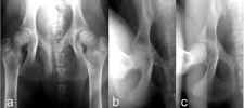
These pictures demonstrate how the PennHIP method discloses hip joint looseness in a dog that appears normal when radiographed using the conventional method [a]. The extended ventrodorsal [a] radiograph, used by the AVA, BVA, OFA and European hip survey schemes is compared to the PennHIP® views that use compression [b] and distraction [c] to disclose and quantitate the amount of hip joint laxity that is present in each hip joint. This laxity information, crucial to the understanding of hip dysplasia, is not available when dogs are radiographed using the standard [a] view.
Despite concerted efforts of organisations like the OFA in the United States, the British Veterinary Association/British Kennel Club (BVA/KC) and other European groups over the last 20 to 30 years to screen for CHD employing the extended hip view, CHD remains common in many breeds.
An improved radiographic method that has been extensively investigated for early diagnosis of hip joint laxity is the PennHIP® method. PennHIP® is a scientific method of evaluating a dog for its susceptibility to develop Hip Dysplasia, and can be used reliably as early as 4 months of age. Coxofemoral joint laxity, which is the first radiographic sign of CHD, may be subtle or absent on the standard extended hip view but is readily detected by the PennHIP® method of radiography.
Hip joint laxity can not be accurately assessed on the extended hip view. Scientific evidence indicates that a laxity index alone can predict the likelihood of future DJD with a high level of accuracy when applied to 16 week old puppies.
Controlled scientific studies have proven that dogs with loose hip joints carry a significant risk of developing joint disease, despite often being classified as normal by the traditional extended (OFA, BVA, AVA) methods. The PennHIP® radiographic procedure involves special positioning of the dog so that the dogs passive hip laxity can be accurately measured. In simple terms, passive hip laxity refers to the degree of "looseness" of the hip ball in the hip socket when the dog's muscles are completely relaxed. Research has shown that the degree of passive hip laxity is an important factor in determining susceptibility to osteoarthritis.
A PennHIP® evaluation results in a confidential report to the owner indicating the dog's Distraction Index (DI). The DI is a measure of passive hip laxity and is expressed as a number between 0 and 1. (A DI near 0 would indicate no joint laxity and very tight hips. A DI closer to 1 would indicate a high degree of laxity and very loose hips.) Dogs with DI scores less than 0.3 do not develop osteoarthritis, with increased incidence of osteoarthritis as the DI increased above 0.3.
Currently, there are more than 50 veterinarians in Australia registered for PennHIP® radiography. The veterinarians at Veterinary Imaging Associates are registered to do PennHIP® radiography. As well, Drs Allan and Nicoll do conventional hip radiography and both radiologists read and score hip joints using the AVA/BVA hip scoring method.
Informative WEB sites:
• PennHIP information
http://www.vet.upenn.edu/ResearchCenters/pennhip/
<< Previous chapter "Application" Next chapter "Breed Average Scores" >>
|
