|
Welcome to a VIA Online Learning Tutorial: This site has been developed to provide learning materials to assist you to make complete and comprehensive radiographic examinations of animals.
Select Chapter:
1. BARIUM MEAL [print this page]
2. ADDITIONAL GIT STUDIES
3. EXCRETORY UROGRAPHY
4. LOWER URINARY TRACT

Traditionally, a barium meal would describe food mixed with barium sulphate being administered to a patient with the intention of observing its passage through the gastrointestinal tract. One situation where barium impregnated food is useful is in the study of oesophageal diseases where one is looking for a stricture, saccular dilatation or diverticulum, as these are not always identified by the usual liquid barium contrast study. Impregnated food may also be preferred to liquid barium when studying oesophageal motility.
Food impregnated barium meals are used less frequently than liquid barium studies for most small animal gastro-intestinal problems.
The animal should be prepared for the study by having had food withheld for a minimum of 24 hours. Ingestion of liquid during this period is permitted. It is preferred that the large intestines be free of faecal material, but it is recognised that circumstances may prevent this on occasions. If the study is to include the large bowel then it is important that action be taken to ensure that the large bowel has been evacuated prior to the study.
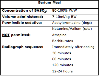
A barium concentration of 80-100% W/W is used. The volume to be administered should be 6-12mL/kg BW. The high dose/kg is recommended for cats and small dogs while the low dose/kg is recommended for large dogs. You can administer the solution into the cheek pouch of the patient using a 50mL syringe with a catheter tip, and allow the patient to swallow it in its own time. It is important to not extend the animal's neck, or to administer the liquid at a rate that is too great for it to be able to drink comfortably.
Make sure you give the patient an opportunity to stop drinking and take breath. If the oesophagus is not to be included in the study, or if you are administering the contrast material to a cat that is fractious, then an oesophageal tube may be used to administer the barium. If sedation is required, acetylpromazine is a useful sedative as it doesn't significantly alter gastrointestinal function. The use of compounds such as atropine, ketamine, and barbiturates will significantly depress gastrointestinal motility, and are not recommended in dogs. Ketamine/diazepam has been recommended for feline sedation.
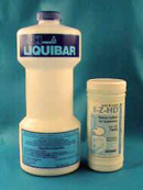
Fig 1: Commercial BaSO4 preparations are preferable to self mixed barium solutions
After administering the barium, take right and left lateral and VD and DV radiographs of the abdomen. Some authorities recommend serial radiography [taking a lateral and VD radiograph on each occasion] at 15, 30, 45, 60, 90, 180 minutes post administration. This is impractical in many cases, particularly in cats. A more flexible regime is to take sequential films after thirty minutes and again after another thirty or sixty minutes. Your decision about the timing of the thirty and sixty minute radiographs will depend upon the rate at which the stomach is emptying and the barium is moving through the small intestines. Then take radiographs hourly until you have identified contrast material entering the large bowel. Usually you will take a final set of radiographs twelve hours after the barium was administered to check complete gastric and small intestinal evacuation of contrast.
The rate of gastric emptying and of small intestinal transit of liquid barium varies greatly among normal cats and dogs. Contrast material may be seen within the large bowel in as little as thirty minutes after administration of barium in some cats but conversely may take as long as six to eight hours in others. As a general rule gastric emptying commences within 20-30 minutes of the meal being administered. In many individuals contrast material will be identified in the duodenum when the first set of radiographs is taken immediately after administration of contrast.
The things you will be looking for when you give an animal a barium contrast study are signs of:
• excessive gastric distension
• altered conformation of the gastric silhouette
• delayed, slow gastric emptying
• segmental or generalised small bowel dilation
• any abnormal mucosal pattern
• normal filling of the large intestine
• complete evacuation of contrast from the stomach and small intestines
The rate of gastric emptying following liquid barium administration is significant if prolonged. If the stomach is dilated, with evidence of peristaltic activity in the body or the pyloric antrum, a gastric emptying time that is longer than 30-60 minutes is strong evidence of gastric outflow obstruction.
Filling defects in the walls of the stomach can indicate infiltrative lesions within the gastric wall. Obvious thickening of the gastric rugae may indicate a chronic hypertrophic disease of the gastric wall. Focal out-pocketing of barium from the lumen of the stomach would suggest the presence of a gastric ulcer.
Within the small intestine the most common abnormalities that you will see following a barium study are signs of segmental or generalised intestinal dilation. Occasionally you will see abnormal mucosal margins, which may indicate inflammatory or infiltrative intestinal disease. A focal, segmental ileus associated with an apple core mucosal pattern is a strong sign of small intestinal neoplasia.
When barium enters the large bowel it usually conforms to the shape of the colon, and ultimately forms the shape of faeces. Radiographic variations in the large intestinal mucosal margin and the diameter of the large bowel following a barium meal are variable, and usually not a reliable sign of pathology. To study the large bowel a retrograde contrast study is used.
Footnote: In cats a combination of ketamine (3mg/kg) and diazepam (0.1mg/kg) was an effective sedative which doesn't affect the gastric transit time.
Steyn P.F. and Twedt D.C. (1994) J. Am. Anim. Hosp. Assoc. 30:78-80.
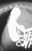 Fig 2 Fig 2 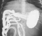 Fig 3 Fig 3
Normal stomach and small intestines 15 minutes after barium administration.
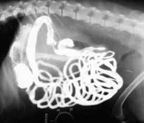 Fig 4 Fig 4 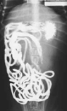 Fig 5 Fig 5
The same dog as figs 2-2, 2-3. These radiographs were taken 45 minutes after barium administration. Barium is visible within the large intestine.
Next chapter "Additional GIT Studies" >>
|