Welcome to a VIA Online Learning Tutorial: This site has been developed to provide learning materials to assist you to make complete and comprehensive radiographic examinations of animals.
The technique of excretory urography allows the renal silhouette to be identified during the nephrogram phase of the contrast study. Contrast nephrography (or excretory urography) is a simple and safe technique which allows identification of the size, shape, location of the kidney and which yields useful information about the homogeneity of the renal parenchyma as well as excretory function of the kidney in question.
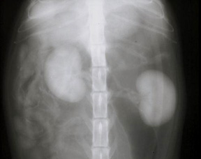
Fig 1 [a-c]. [click to enlarge] During the nephrogram phase [a] renal arteries and veins are visible and often there will be good corticomedullary distinction. The excretory phase [b and c] reveal gradual loss of renal opacity and opacification of the renal pelvis and ureters.
The principle of excretory urography is to inject intravenously a water soluble radiopaque substance which is selectively excreted by the kidneys. The choice of contrast is simplified by the fact that most of the iodine containing water soluble contrast material currently available are renally excreted. Pharmacological products in this category may be formulated as ionic or non-ionic preparations. In general, either class is effective for the purpose, but non-ionics are generally more expensive that the older ionic preparations. In Australia media such as Urographin (76%), Urovison (58%) or Omnipaque (300mgI/mL) are readily available. The dose rate for intravenous administration is 880mg of iodine per kilo of body weight. After intravenous injection the material circulates systemically throughout all body systems. Their plasma half life is relatively short, and they are selectively concentrated and excreted via the nephrons. Maximum renal parenchymal opacification occurs approximately 60 seconds after the injection of a bolus of contrast medium, and radiographs taken at this time are referred to as being of the nephron phase, or a nephrogram.
The study:
[1] Patient preparation: Nil by mouth for 36 hours prior to radiography and ensure evacuation of the large intestine prior to commencement. The study will be easier to do if the patient is anaesthetised.
[2] Survey radiography. Take survey radiographs of the entire abdomen. The purpose is to establish:
• normal non-contrast radiographic features of the abdomen
• adequate exposure factors for the study
• that faecal material won't interfere with visualisation of the [entire] urinary tract.
[3] Place a venous catheter in the cephalic [preferably], or the saphenous vein and tape it in place. An 18-20g catheter will simplify rapid injection of contrast medium and reduce the possibility of [harmful] extravascular injection. Catheterise the urinary bladder and drain the urine. Retrofil the bladder with gas and leave the catheter in place. Some radiologists prefer to use a ballon tipped Foley catheter but a snugly fitted urethral catheter will usually suffice to prevent leakage from the lower urinary tract during the procedure. A three way stopcock attached to the catheter will help retain gas in the bladder as well as facilitate further drainage of urine from the bladder during the procedure.
[4] Positioning. Several sets [about 4] of ventro-dorsal and right lateral radiographs of the abdomen will be taken over a 15-20 minute period. The use of positioning aids [foam trough, sandbags] will assist in ensuring adequate positioning for each radiograph. While the procedure can be done on a conscious or sedated patient, anaesthesia eliminates personnel exposure while enabling rapid and precise repositioning during the procedure. Start with the animal positioned for the ventro-dorsal view.
[5] The injection and nephrogram phase radiographs: Use warmed [to body temperature] contrast material. Warming the contrast agent reduces its viscosity and facilitates rapid injection of the bolus of liquid. Having made these preparations, make a rapid injection of contrast material and expose the ventrodorsal radiograph 40 to 60 seconds after completion of the injection. Quickly reposition the patient for the lateral exposure and take it as soon as possible after the ventrodorsal radiograph. This will enable two radiographs of the abdomen to be taken between 40 and 90 seconds after the injection of contrast. The purpose of this first set of radiographs is to allow visualisation of the renal parenchyma.
[6] Subsequent radiographs: During the next five to 20 minutes the kidneys will concentrate and excrete contrast material, allowing visualisation of the renal pelvis, the ureters and the urinary bladder. Subsequent radiographs should therefore be made at five and ten minutes post injection. The ureters, the ureterovesicular junction and the bladder will be studied at these times. The bladder study will be a double contrast one.